


Geoff’s Narrations
The GIST
The Blog
THE GIST
- Many studies suggest something is up with the mitochondria in diseases like chronic fatigue syndrome (ME/CFS) and long COVID but the question remains: if the mitochondria are not functioning well how did they get that way? While we don’t have the answer to that, recent studies are providijng some clues.
- When muscle tissues (apparently from healthy controls (?)) were exposed to serum from ME/CFS and long COVID patients for 48 hours, the muscles switched from producing energy using the mitochondria (i.e. using aerobic energy production) to producing energy anaerobically.
- High levels of mitochondrial and non-mitochondrial oxygen consumption capacity indicated that the serum caused the mitochondria to work really hard. As time went on, though, the mitochondria began to fade and break up and the muscle tissues became highly fatigued.
- The authors hypothesized that a “stress-induced hypermetabolic state” ultimately resulted in “severe deterioration of muscle function”.
- These are not the first authors to suggest that an initial burst of hypermetabolism permanently affected the ability of ME/CFS and long COVID patient’s cells to produce energy.
- Earlier this year a study examining the effects of the Tick-borne Crimean-Congo hemorrhagic fever virus found that energy demands soared in the early stages of the infection – again creating a “hypermetabolic state”.
- Some of the patient’s mitochondria wilted under the stress and began to produce energy using the anaerobic energy pathways to generate energy. Thirty days later – long after the pathogen had been vanquished – their immune cells still remained in a state of “metabolic insufficiency”.
- Similarly, several studies suggest that the energy production systems of B-cells have become ‘overstressed” in ME/CFS. This appears to prevent many B-cells from maturing – leaving an important part of the immune system hobbled when fighting off pathogens.
- Indeed, Avindra Nath has proposed that a lack of B-cell maturation may be the primary driver causing ME/CFS.
- Other studies have found evidence of exhausted and overly stressed T-cells that could be tied to an inability to produce sufficient energy.
- All in all these studies suggest when the mitochondria of people with ME/CFS and/or long COVID are put into a highly stressed state such as during an infection, they become exhausted and are not be able to recover.
- Bob Naviaux’s Cell Danger Response model proposes that the hypermetabolic to hypometabolic switch is operative in ME/CFS. He believes our cells become supersensitive to danger signals such as ATP when it is released from a cell when it becomes stressed.
- He believes that antipurinergic drugs like Suramin will ultimately be able to return both the cells and the patient to health.
- Once again the hypersensitivity theme shows up. From the overly sensitive glial cells in the brain to the hypersensitive pain processes found in the central nervous system, the spine, and nerves in fibromyalgia, finding ways to calm things down will surely play a role in overcoming these complex diseases.
Recent studies provide some clues. The latest came from a fascinating abstract “571P Muscular metabolic plasticity in 3D in vitro models against systemic stress factors in ME/CFS and long COVID-19” from the 2024 Neuromuscular Disorders conference (thanks, Jutta for the tip! With its focus on the mitochondria and the muscles this Spanish study really hit some hot spots.
When muscle tissues (apparently from healthy controls (?)) were exposed to serum from ME/CFS and long COVID patients for 48 hours, a bunch of bad things happened.
The fact that the serum from both the ME/CFS and long COVID (LC) patients produced the same result suggested that the kind of trigger (SARS-CoV-2 virus – LC; multiple possibilities – ME/CFS) didn’t matter – the same core dysfunction was present in both. It brought to mind Akiko Iwasaki’s admonition to stop focusing so much on the spike protein in long COVID and look to core abnormalities that pervade all these diseases.
The ability of the muscle to contract; i.e. produce force (muscles produce force by contracting) was “severely compromised” suggesting that these muscle tissues were probably pretty much dead in the water.
At the same time that was happening, enhanced levels of glycolytic enzymes suggested that the muscle tissue’s ability to generate lots of energy via the aerobic energy production pathway had pooped out – causing them to rely on the less efficient anaerobic energy production.
At first glance, the myotube hypertrophy (increased incidence of immature muscle cells) found suggested that muscle cells were simply gearing up for activity. The fused mitochondrial networks, though, suggested that adding the ME/CFS/Long COVID serum to the muscle tissue put the mitochondria under severe stress and the myotube enhancement was a compensatory mechanism.
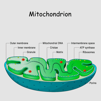
Serum from ME/CFS and long COVID patients caused the mitochondria in the muscle cells to become highly stressed and ultimately dysfunctional.
Indeed, both mitochondrial and non-mitochondrial oxygen consumption capacities were elevated – suggesting that the serum caused the mitochondria to work really hard. As time went on, though, the mitochondria began to fade, and break up and the muscle tissues became highly fatigued.
The authors hypothesized that a “stress-induced hypermetabolic state” ultimately resulted in “severe deterioration of muscle function”. In this scenario, something in the serum caused the muscle cells to get revved up – and exhaust themselves – resulting in the exercise intolerance in ME/CFS and long COVID.
An “Energy Maladjustment”
These are not the first authors to suggest that an initial burst of hypermetabolism permanently affected the ability of ME/CFS and long COVID patient’s cells to produce energy.
Earlier this year a study examining the effects of the Tick-borne Crimean-Congo hemorrhagic fever virus found that energy demands soared in the early stages of the infection – again creating a “hypermetabolic state”. The ATP upregulation, the turn towards an increased use of amino acids, and the increased fatty acid oxidation suggested the immune cells scrambling to keep up the fight against the virus.
That made sense. We know that during an infection immune cells have to dramatically rev up their engines.
There was a catch, though. Some of the patient’s mitochondria wilted under the stress and began to produce energy using the anaerobic energy pathways (glycolysis/pyruvate metabolism) to generate energy. These patients had a more difficult time fighting off the virus – indicating that the ability to produce energy plays a crucial role in being able to fight off the virus.
Then came the post-infectious clincher. Thirty days later – long after the pathogen had apparently been vanquished – their immune cells still remained in a state of “metabolic insufficiency”.
Indeed, the vast majority of patients (83%) were experiencing fatigue strong enough to inhibit their daily activities leading the authors to conclude they were in a postviral fatigue state. Besides fatigue, musculoskeletal pain (75%), anorexia (50%), weight loss (50%), headache (38%), palpitation (38%), and sweating (38%) were found.
The authors declared that an “energy maladjustment” had occurred and concluded that “metabolic rewiring during the recovery phase potentially leads to postviral fatigue”.
Immune Cells
The B-cell Bust in ME/CFS
Several studies suggest that B-cells’ energy production systems may have also become ‘overstressed” in these diseases.
Studies that have been tracking down why so many “naïve” or immature B-cells are present in ME/CFS have found that these B-cells have also turned to anaerobic energy production. The lower levels of mitochondria and the higher levels of lactate associated with them provided a reason why these immature B-cells may be having trouble making it to maturity.
These immature cells have also been associated with a breakdown in autophagy; the ability to safely recycle the cells contents including the mitochondria. Autophagy-deficient cells are associated with, guess what, high levels of inflammation. The Simmaron Research Foundation has found evidence of autophagy problems in ME/CFS.
Since antibody-producing B-cells play a major role in fighting off infections, having a bunch of immature B-cells hanging around the immune systems of ME/CFS patients would not be much help.
Interestingly, Avinda Nath believes immature B-cells constitute “the primary defect” (in ME/CFS). The inability of these cells to mature leads to “immune exhaustion and activation of innate immune responses”; i.e. the adaptive (later) immune response that the B-cells participate in punks out leaving the early and more inflammatory innate immune response to kick in.
Note that these studies in these different cell types appear to duplicate the same energy transition that exercise studies indicate has happened: a broken aerobic energy production turns to the less efficient and dirtier anaerobic energy production.
Could it all have begun with overstressed mitochondria?
T-cell Troubles
Vishnu Shankar’s ME/CFS Stanford study (unpublished) suggests it might have. He found that overstressed mitochondria in T-cells were breaking down and releasing high levels of free radicals/reactive oxygen species. Those high levels then damaged the mitochondria – causing even more oxidative stress – and throwing people with ME/CFS into a vicious cycle.
Liisa Selin might agree as well. She found exhausted T-cells in ME/CFS were associated with increased levels of oxidative stress. That made sense given that damaged mitochondria produce a lot of oxidative stress. In a preliminary study she found that a nebulized agent with antioxidant was able to help restore T-cell functioning.
Overtraining Syndrome
While we don’t know if a hypermetabolic state precedes it, it appears that overtrained athletes are stuck in a hypometabolic state as well. Metabolomic studies suggest they are in “procatabolic environment”, in which their muscles are being broken down to provide energy.
The authors proposed that the “global reduction in metabolic pathways” found, may have been produced to save energy and redirect energy during a state of (energy) starvation; i.e. it suggests that the athlete’s mitochondria, for whatever reason, became overstressed and were unable to provide normal amounts of energy.
Bob Naviaux believes the mitochondria in ME/CFS patients become supersensitive to signs of cellular stress. Anti-purinergic drugs like Suramin could help.
Bob Naviaux’s Take
Indeed, a state of energy starvation or hypometabolism – perhaps akin to Naviaux’s Dauer state where the cells shut down the metabolism – has long been proposed to be present. I asked Bob Naviaux whether this hypermetabolic to hypometabolic switch fit his Cell Danger Response hypothesis and it did.
Yes. Everyone experiences a transient hypermetabolic state with the beginning of CDR1 and fever during an acute infection. Most people experience this dozens of times in a lifetime. 90% of the time this resolves without any consequences after a few days to weeks, and we recover completely.
Only about 10% of patients have lingering symptoms that last for more than 6 months. In the patients who develop ME/CFS, Long-COVID, and many other hypometabolic, multi-system, chronic fatigue syndromes, mitochondria and cells enter a chronic, but reversible physiologic state that protects the patient and cells from new injuries and chronic threat, but at the expense of a dramatic decrease in functional capacity. This occurs because the chronic response to danger (the 3 phases of the CDR) siphons energy and resources away from baseline health for purposes of cellular defense.
Our research suggests that this state is maintained chronically by receptor-mediated hypersensitivity to ATP-signaling. Very small stresses can trigger setbacks (PEMs) because the body is hypersensitive to small amounts of eATP release that occurs with stress or exertion.
I believe that when ATP signaling sensitivity is restored to normal by a course of suramin treatment, or eventually with other antipurinergic drugs the might one day be taken by mouth, that full recoveries from ME/CFS, Long-COVID, and related disorders will be the norm and not the exception.
Conclusion
A hypermetabolic state that overly stresses the mitochondria early on could be setting the stage for a long term case of hypometabolism; i.e. reduced energy production in diseases like ME/CFS and long COVID. Why the mitochondria might be failing this way in people with these diseases is unclear. Bob Naviaux’s research suggests that the cells have become hypersensitive to stress factors (eATP) produced by the cells and he believes that anti purinergic drugs will ultimately be game-changers.
Once again we see the hypersensitivity theme show up. Jarred Younger believes the glial cells in the brain have become hypersensitive to stimuli in these diseases. With hypersensitive pain processes present in the central nervous system, the spine, and the peripheral nerves in the body, fibromyalgia, of course, is the queen of the hypersensitivity diseases. What’s causing such a twitchy state is unclear but finding ways to calm things down will surely play a role in overcoming these diseases.


Nice article.
Look forward to hear about next steps with the nebulized agent when you do your blog on Nancy Klimas 👍
Is suramin being trialed?
The suramin trial has been trying to get going for a long time – it’s just never made it to the start line. It reminds me of Dr. Klimas’s TNF-a/mifepristone trial – it’s always seemed about to start!
I asked Dr. Naviaux about it but have not received an answer yet.
yes, so sad for us with the sumarin, it is awfull… Nancy klimas, for ages wants to do things but it never happens. and Naviaux does not give an anwer (yet). And does he do something with it? trials hardly needed? hopelous. decades of fals promisses/hopes and so many of us really can no more, like me…
Yes I don’t know why I even bothered asking
There’s a few researchers that one can put little faith in. Despite earlier promise, Younger is another I now doubt. Trialling all sorts of botanicals, it all feels scattergun
I agree it’s not THAT exciting but he is pursuing things that prior research suggests could help tamp down inflammation. He’s actually quite selective. Would I rather he run a trial of some hotshot drug? Yes, but I imagine that’s really hard to get going.
I would say failed attempts. Other than people with ME/CFS I can’t imagine that anyone wants to get that trial done than Naviaux! If it succeeds not only does he get to help a lot of people but it validates his validates his hypothesis – which he worked on for many years.
Cort,
are you able to comment on the following? :
Functional medicine doctors in different arenas than those featured on Healthrising have suggested that a lack of mitophagy (destruction/replacement) of damaged mitochondria slows down or halts recovery from CFS.
I am wondering whether restoring mitophagy has the potential to halt excessive purinergic signalling.
I would like to comment on the hyper vs hypo metabolic states mentioned here:
As you know, there are a few types of heat generation possible by cells: that from exercise and that without exercise. Without exercise, there is either shivering or non-shivering heat generation. There are obviously various phenotypes required for mitochondria to successfully do their jobs. If mitophagy were to fail, then mitchondria could remain in action that are not suitable for recovery.
We know that in a fever state the mitochondria will work hard to produce heat but not work hard to produce energy for things like ribosome activity so that they do not aid virus or bacterium replication.
In Rob Phair’s research we also saw that the Itaconate shunt may be playing a role in depriving the cell of adequate ATP production via normal aerobic glycolysis. I think I mentioned it here before, but , the purpose of Itaconate and the Itaconate shunt is to:
produce more cytokines (and possibly chemokines) to fight microbes.
So in that state:
1. The mitochondria are not moving on to recovery because they are stuck in the poisoining state — poisoning attackers.
2. the mitochondria are working hard, but not on ATP for exercise, only ATP for itaconate, and for eATP purinergic signalling.
It seems to me that restoring mitchondrial autophagy can correct this state.
I acknowledge people’s comments below that the calcium ion channel is blocked in CFS. I assume they mean in T-cells. I believe that you treated that topic with regard to T-cells. I do not believe that muscle cell calcium ion channels are blocked since I do believe that
after hypoxia from hypoperfusion, when there is incorrect Renin-Angiontensin-Aldosterone function in vessels near muscles, Ischemic Cascade occurs, and that that includes bringing in additional calcium when Glutamate excitotoxicity occurs outside the cell membrane. I do not see a calcium ion channel failing there.
Several small studies have suggested that autophagy is impaired in ME/CFS. I don’t know if restoring it would help with the purinergic issue.
Thanks for the overview of itaconate shunt 🙂
I’m sorry, Cort, I should have called it mitophagy since it is different from whole-cell autophagy. The cell ought to replace damaged mitochondria, or mitochondria which are the wrong phenotype for normal function if they have been “configured” for low function as regards exercise.
Bob Naviaux explained that the Cell Defense Response, CDR, causes a change in the expressed phenotype of the mitochondria. I think it would be beneficial to induce mitophagy without attempting to induce autophagy.
“When muscle tissues (apparently from healthy controls (?)) were exposed to serum from ME/CFS and long COVID patients for 48 hours, a bunch of bad things happened.”
This suggests a transmissible agent and most likely the same agent since all kinds of different infections wouldn’t necessarily cause the same thing.
Now let’s explore the B cell and T cell findings.
In 1986, two researchers working in the Gallo Aids lab discovered a new virus. At first, they had a hard time keeping it alive because it killed all the cells they put it in. When they resolved this, they found that it was killing B cells, so they named it HBLV (Human B Lymphotropic VIrus}.
Further research found, however, that it was also killing T cells so for reasons I have never understood, they changed the name to HHV-6 and then subdivided that into HHV-A and B.
HHV-B is a common cause of childhood roseola and is in about 90% of the population. A test at Quest Labs will tell you if you have HHV-6 B.
This is not true for HHV-6 A which must be tested for through a specialty lab at a high cost.
“According to the CDC, molecular analysis showed a higher prevalence of HHV-6A but not HHV-6B or HHV-7 in CFS patients (64,101,102), and HHV-6A could also be isolated from these patients (103). Whether this reflects an association or the consequence of an immune dysregulation remains to be determined.”
There are scientists who believe that HHV-6 A is not a HHV-6 variant, but an entirely separate virus. One that we are not testing for. Why?
I believe Dr. Klimas is finding more HHV-6 reactivation than other herpesviruses – I don’t know if she’s looked at types or not. It puzzles me, as well, as this seemingly rather simple question – are these different viruses – has not been cleared up yet. I guess it’s not so simple after all.
Cort, in addition to Prusty’s Sep 2024 HHV-6 and mitochondria presentation video linked above in reply to Betty Mekdeci, you can also find a German language script of the presentation here https://www.fatigatio.de/fileadmin/Projects/07_fatigatio/user_upload/Fachtagung_2024/Fachtagung_2024_Uebersetzung_Prusty_NEU.pdf, which you could enter into DeepL for translation.
Hmm. I guess the CDC knows how to test for HHV6-A
“According to the CDC, molecular analysis showed a higher prevalence of HHV-6A but not HHV-6B or HHV-7 in CFS patients (64,101,102), and HHV-6A could also be isolated from these patients (103). Whether this reflects an association or the consequence of an immune dysregulation remains to be determined.”
About what you write, Prusty (who researches HHV-6 in conjunction with ME/CFS) found little communcation miRNAs as well as viral proteins (dUTPases) by which the virus manipulates cells and mitochondria. I think he said the same may apply to other viruses, and has found them for EBV and HSV-1 too.
He gave an English language presentation on “Mitochondrial dysfunction and altered metabolism in ME/CFS”, summarizing his results so far, in September 2024 here: https://www.youtube.com/watch?v=YUnBWvw3wGA.
This article had me opening multiple research tabs as I went through and I found this paper detailing some antipurinergic herbal treatments. One of them is Ligusticum wallichii. I had found great relief last year from Ligusticum porterii (Osha) which is a Native American alpine plant known to cure Lyme for some people.
I ended up perhaps taking too much and after feeling great for a while I came down with a candida skin infection thought to be induced by this natural antibiotic disturbing the bacteria-fungal balance in my biome. The problem of course is that when you experiment with herbs you can’t do so under an MD’s guidance. But I might try again at a lower dose. I had assumed Osha worked because it was antibiotic but perhaps it was also antipurinergic… (?)
https://www.ncbi.nlm.nih.gov/pmc/articles/PMC3817725/
Yes osha I had success with.
An indigenous friend from the Rocky mountains was able to get me some.
It worked great as a tea so I decided to try chewing the root whole. It actually split my tounge in several places but probably because i chewed it in my mouth for several minutes as opposed to chew and swallow.seems pretty powerful and spicey
It’s use primarily as a lung
Decongestant.
ill try anything once to rid myself of this horrible disease
How interesting! I would assume taking a probiotic along with the OSHA may prevent Candida. I have an herbalist I will enquire about this herbal and dosing.
Would pine needle tea perhaps help ? I searched sources of suramin and that came up .
Thanks! Someone suggested the herbal Osha, so am looking into that.
Like Sonya Gradisnek, head ME scientist of Griffith University Australia, says in her you tube interview with the Sweden guy, If the calcium ion channels are closed, which has been identified in All CFS patient supplied blood, Inflammation happens throughout the body in all ways, these channels are everywhere, and concentrated in certain important areas like the brain, heart, spleen, muscles etc
And the Eyes and ears, Now I have no more eye and ear infections, a basic life, with light walking, exercise, yoga, chores, by using 7mls of naltrexone every 2 hrs to keep calcium ion channels opened. People underestimate its importance, but look at Kalemic Periodic Paralysis, which my son and his father have ,without potassium they become Paralysed!! The key blocks of Sodium, potassium, magnesium and calcium are essential for good health!
All viruses enter through calcium ion channels and in susceptible people cause them to close . Naltrexone restores this by blocking the oipiod receptor which the opens calcium ion channels.
may i ask, do you mean ldn? or really naltrexone? for ME/cfs? i am so so bedridden in dark room, still decline and still inflamed (up and down)
Hi Kunjin, low dose naltrexone is just naltrexone, I dilute a 50mg tab in 50mls of water, then dose with a 1 ml syringe, I had to start with .1 of a ml, so 1 drop and titrate up slowly, I am the first person to use it unconventionally, so every 2 hrs, as calcium ion channels close again after that, it all depends on how much you are using, naltrexone is not toxic until 800mgs a day, I use about 80mgs over a day, always proceed with caution, especially if you are so severe, I find some caffiene helpful too, it helps calcium influx, raises noradrenaline and increase Cerebral Spinal Fluid. Sorry you are so unwell, I remember that! I have some followers that it works for too. It’s safer to do most movement in the first hour. All the best.
may i ask how you tapperde up to 80 mg and for how long, verry verry slowly?years? or so and how divided? thanks!!!
can you sleep with it? read many people must take way less in the morning..
Like walking etc, in the first hour. I can stand now all day if i wish, and have zero fatigue, pem, pots or pain. I do tend to do too much at times though, lol which can bring some of that back, but keeping up the ldn helps immensely.
thank you so much for giving me hope and the effort of writing!!! must keep it short because verry unwell…. glad you are so much better!!! 🙂 now an online gp finding in neighbourcountry if i can and if gp is willing. xxx!!! please stay wel!!!
Thankyou I shall, as I said this is breaking stuff, if you can get the 50mg tab and dilute it you’ll be able to titrate easier and play with it more, morn and night at first, start very low and titrate slowly, don’t forget caffiene too if needed, you can download the precribers info from Google for yr doctor, just type in ldn prescribers information. There are ldn, cfs fb groups too, but I’ve left them as the admits are closed minded, it’s mostly only prescribed once daily, but for the majority dosent work. Here is my email if you need anymore help, vgray1221@gmail.com
thank you so much sweety!!!
first finding a gp in a neighbourcountry f willing to prescribe. it is so difficult from bed. and they are not good in listening, can not say give me 5 mg pill. they have there own opinion and maybe i even do get none… please cross your fingers for me!!!xxx!!!
Konijn, there is an apothecary in Germany specialised on low dose naltrexone (LDN= https://www.low-dose-apotheke.de/home/ , info(at)cityapotheke-goettingen.de . If you can get a prescription, you could ask the apothecary if they also ship to your country. They do low dose liquid as well (Tropfen – drops) so you can start very low and do not need to dissolve tablets yourself.
But if you are so severe, I’d recommend you start very low and get drops with a low concentration, so you can start at 0,1mg like Vanessa did or even less. I suppose that apothecary can do any concentration.
I started on LDN 0.5 mg for 2 days and had strong initial side effects so I needed to stop, and will need to start again with a much lower dose later. If you start it, do so very carefully because side effects also changed the perception of my energy limits, which made me overexert and crash.
For me it is known that I have strong reactions to many medications at low doses and have multiple chemical sensitivities, so if you are similar to that, it could be a sign you also need to start very carefully.
And as Vanessa said, her dosage scheme is new and unconventional, so you may want to start LDN the traditional way first, e.g. very carefully titrating up from a very low dose, but only once a day, and remaining at one dosage level until the body has adjusted, only then trying to go up (usually not higher than 4.5 mg/day but in 1 dose).
You can show the GP the following 2 publications and say that Low Dose Naltrexon (LDN) it’s one of the most well-known off-label medications for ME/CFS which helped 70% of patients in this 2019 finnish study https://www.tandfonline.com/doi/full/10.1080/21641846.2019.1692770 and also helped in these 2020 case reports https://pmc.ncbi.nlm.nih.gov/articles/PMC6954765/ . If your GP speaks German, you can make a screenshot of minute 34:08 of this Charité Berlin medical training presentation for usual LDN dosage scheme. Good luck!
Have you tried nicotine patches yet? https://www.healthrising.org/blog/2023/12/07/nicotine-patch-long-covid-chronic-fatigue-fibromyalgia/ After I had gotten used to this medication, they help me recover from crashes. I use the Sefudun patches from Amazon, which I can cut into any dose.
By “very carefully titrating up from a very low dose, but only once a day, ” I mean taking only one dose a day.
Here’s the video link to the Charité presentation: https://player.vimeo.com/video/917863881 min 34:08.
thank you verry much for the link!!! i ask you the same question as Vanessa. no need to answer if you do not like it!!! where you bedridden to? i am 30 years ill and pretty severe…afraid it will not help. and how do you know it moves the immunesystem in the right direction. i was given in 2001 for a half year 3 tupes of antibiotics. was allready severe but could walk into the hospital. after 2 weeks i was bedridden. everytime if they did something with my immunesystem i declined.. thanks!!!! hope you mprove further!!!xxx
thank you so much!!! i am so sorry you crashed with 0,5 mg verry verry hard! and need to stop and then to start all over again!!! are you back on your previous aseline now? i really do hope so! wich side effects where it if i may ask. if you do not want to write about then, you ofcourse must not!!! thanks verry much for the many links but my gp is against medication and in my country it is still get and cbt. it would be a miracle if i could get ldn from 1 gp in a neigbourcountry (after first paying 900 and Euro’ for on-line examination and if he even is wanting to prescribe it to me. no, i did not tryd the nicotine patches yet. i hope i can “make it” to the neigbourhood country gp and he will prescribe ldn for me first. i need a miracle… wich screenshot from charité berlin please? but most of all, cross your fingers for me that i get the ldn and that it helps. me and many others, all of us…
– For the German LDN dosage screenshot, open this video on your computer https://player.vimeo.com/video/917863881, open the video at minute 34:08, and then make a screenshot of it using your computer keys (Fn key + print key), or take a photo of it with your mobile phone.
– send an English email to the LDN apothecary I recommended info(at)cityapotheke-goettingen.de and try asking them if they know from which doctor you could get a prescription.
– In your place, I would really consider giving nicotine patches a try. They are cheap and you can buy them without prescription on Amazon (for example Sefudun quit smoking nicotine patches, which look like these: https://www.amazon.sa/-/en/Smoking-Patches-Delivered-Hours-Patches/dp/B0BL7RNMYG ), or in apothecaries. – When it is a thin matrix patch like the Sefudun patch, it can be cut into any dose. (Old-style reservoir patches must NEVER be cut, so make sure you have a matrix patch – which most modern ones are.) For me, there were much less initial side effects from nicotine than from LDN. – It works really easy – you do it it rounds: e.g. in the first round start with a low dose e.g. 3.5 mg for a couple of days, then pause for a couple of days, then do another round and so on. If you apply a very small piece of patch, stick a normal band-aid over it to keep in place. Initially nicotine can be very activating so the most important thing is to rest while applying it so you don’t crash; over time it can become more calming in my experience. It can raise heart rate so I think project safety guidelines https://drive.google.com/file/d/1v8UgRJ4aafrs_RoKRHQKfhUxT8O7VVYQ/view say to be careful if you have cardiovascular issues .
Here’s a short how-to for the nicotine patches: https://drive.google.com/file/d/1jJ4TzTPjuJaJjEd9VPADW1BcbhbFOxuP/view . The project manager also answers email questions.
– The LDN side effects I think went away after a couple of days after I stopped it. I am not bedridden but severe. Sorry I can’t answer more about side effects, do not have more energy! Best wishes!
I mean not completely bedridden.
Hi JR, that is why I use the ldn unconventionally, every 2 hrs, this stops crashes, pem, fatigue,etc. You will be amazed how well it works like that!
One problem with it is the body becomes tolerant to it, and you constantly need to titrate up, I cannot now at 7mls, still works, but not as long or powerfully. I get enough naltrexone by pretending to be a alcohol needing it. All the best
thanks so much!!!xxx!!!
I got LDN (low dose naltrexone) from a compounding pharmacist. I’m very med. sensitive so started with 1 mg and working my way up. Find a compounding pharmacy, if you can, and have them make it in any dose you decide to start with.
They may be able to mail it to you. That’s what I do.
I bought it from a compounding pharmacy which mailed it to me – it’s worth a try – as is red light therapy – a blog is coming up on that. 🙂
Hi Cort, may i ask you (no need to answer) on what dose you are? how you build up? wich effects it has or side effects? shurelly no need to answer…!!! thanks!!! feel safer when more peolple take it… xxx!!!
Looking forward to learning about red light therapy, I’m guessing it doesn’t mean getting a ticket for running a red light 🙂
Sorry, I couldn’t resist. A little humor never hurts.
thanks verry much!!! but first the big problem here for a prescription. may i ask on what dose you are now and over how long time build it up? and wich side effects or good effects? you must ofcource not answer that!!! I just wonder if there are people getting worse with it… xxx!!! good luck to you!!!
Hi Konijn,
I don’t know where you are or what your situation is but I got my primary care physician to prescribe LDN. She didn’t know much about ME/CFS but was willing to work with me when I told her about LDN. Not all doctors are. Some of them don’t believe us when we tell them.
I took the 1 mg dose for 2 months and didn’t notice any change at all, either good or bad. I am now on 1.25 mg dose- for one month – and am seeing slightly more energy, with the increased dose, and also less POTS. I haven’t had any negative side effects at all.
Good luck to you, too! 🙂
Hi Susan, thank you verry verry much!!! I live in Belgium where it is still get and cbt… my gp would never prescribe it for me, here it is in fact unknown. Also mestinon or low dose abilify. I am severelly bedridden. GP is also not willing to learn about it and off label meds is shurely a no go… I even do not get meds for excample for slow gastric amptying, pain, etc It is crazu that 1 mg did nothing for you but a tiny increase to 1.25 mg and you are seeing slighly more energy and less POTS! i am glad you did not had any side effects! I hope, with maybe incresing more, you will see even better results!!! good luck with it!!!xxx!!!
Hallo Konijn, groetjes uit België.
I ask a psychiatrist to prescribe me a box of LDN 50mg, and the pharmacy dilutes it into 4mg pills that I take daily. I started at 0,5mg though, and had some slight side effects the 1st two weeks. I definately have my life back, but I was a milder case, only with severe episodes of a month. Ik wens u het beste Konijn !
Konijn, I wonder if you can order from here: https://www.buy-pharma.md/Naltrexone-p-360.html
You don’t need a prescription to order from there
Hi Rebekah,
what i read is that you need a prescription and must see by yourself for country rules. the prescription sttos somewhere below. (to ill to remember but looked at it). it is also normal because it is a med and must go through many countrys. i even do not know if they are thrustworthy. but thanks verry much for the effort!!!xxx!!!
Hi Konijn, I’ve ordered many medicines from them without a prescription, and have found them very trustworthy. The shipments have always come through all customs to me in the USA. Maybe worth a try for you.
may i ask you one more question please? no need to answer if you do not like it!!! where you bedridden to? i am 30 years ill and pretty severe…afraid it will not help. and how do you know it moves the immunesystem in the right direction. i was given in 2001 for a half year 3 tupes of antibiotics. was allready severe but could walk into the hospital. after 2 weeks i was bedridden. everytime if they did something with my immunesystem i declined.. thanks!!!! hope you mprove further!!!xxx
Hi Konijin, yes at first for a few years, I had ebv that was my trigger, as JR said titrate slowly only going up 1 drop every 2 weeks at first, I have had a few times where I’ve improved slightly enough to drop etc, I was raising 2 children then too, so had to do stuff but it wasn’t easy, recently crashed before ldn by truing to work and menopause plus the cfs of course. Ldn can be a bit tricky, and it’s not the full answer but it definitely helps me. So I can live basically again, which I’m grateful for it as being bedbound is hard I know, and im sending you good luck and hope 💜 don’t forget some caffiene if needed too.
Sorry that wasn’t very clear, typos, I’ve only been using ldn 2 yrs, and had cfs for 9 years, I was saying, bedbound about 2 years then improved to care for my kids, somehow, I did every type healing I could find, caffiene helped but was still crashing alot. All the best
hi Vanessa, thaks for your answer! so glad you improved!!!so that you can basically live again and take care of your children. yes, being bedbound is verry hard!! so glad you could escape from that!!!xxx!!!
If you want, I can give you some online pharmacies within Europe that can send it to you quite safely.
Although never a guarantee.
thank you verry much!!! but i need firs aprescription and that is the problem… xxx!!!
Your welcome 🙏 Take care and Best of Luck and Blessings 💓
thank you!!! xxx!!!
Klaus Wirth believes damaged calcium channels play a major role as well.
https://www.healthrising.org/blog/2023/02/22/chronic-fatigue-syndrome-calcium-muscles-startup/
thanks Cort! as allways!!!
🙂
I love reading the comments to see everyones ideas and contributions!
I’ve been pondering metabolism lately in regards to my own circumstances.
I’ve been listening to Dr Mark Donohoe (Australian GP who I’m booked into see next year) talk about the state of hypometabolism in ME/CFS and he likens it to animals in hibernation, even pondering whether we are ancestrally designed for a slower period each year, going into keto when carbohydrate supply and stores ran out, and whether our modern lifestyle are disrupting and stressing that natural balance. I do feel that the normal western lifestyle is over stimulating and I’m trying to go against the grain and choose a life that is slower and calmer.
Dr Donohoe also noted that many of his patients have low body temperature as a reflection of the hypo state and indeed, I’ve been checking mine each morning and I run low. One morning a couple of week ago I was at 34.5C, which is mild hypothermia, and given I live in the subtropics and it’s spring, this is obviously a bit abnormal. However, it reflects my experience of being freezing cold to the point of not being able to function in winter (even in the subtropics).
Another hypothesis around this I have been exploring recently is how these hyper and hypo states may tie in with polyvagal theory. I’ve recently discovered the Primal Trust course (worth having a look if you haven’t already Cort) and Dr Cathleen King connects the polyvagal dorsal vagal shutdown (ANS freeze state) to Naviaux’s cell danger response. I think it’s an interesting hypothesis and I can certainly relate to it from my personal experience of long term chronic stress and a very dysregulated nervous system coinciding with many of my fibro and CFS symptoms.
As always, I continue to ponder if stress and my psychology alone caused my biology to shift, or if infection/toxicity and depletion caused my nervous system to become sensitised. In my case I can see how both happened at the same time creating the perfect storm. I’m slowly working my way out if and a big focus for the past few years has been supporting and improving mitochondrial health and metabolism. I’m working out at the gym 4 x a week without crashing now, so I’m doing something right. But it takes a lot of strategy and effort round the clock to manage. I’m about to start Primal trust as I feel that body of work is ideal for my next stage of recovery. And then hopefully Dr Donohoe can help me with the last stretch next year.
That theory of Dr Donohoe’s is very intriguing and Id like to read more. My husband’s ME/CFS symptoms worsen terribly in the winter, making him basically bed and couch bound that season. Come spring, symptoms improve, he struggles again in July with the humidity and then perks back up come fall. His MD is perplexed also. We have actually (sadly) commented that his winter hibernation is coming….it prevents any winter travel or outings.
To tease out the pathobiological matrix, the authors of “571P Muscular metabolic plasticity…” would have to do two things:
a)
repeat the same experiment (same patient group), but this time with serum harvested at two different time points:
once during clinical “baseline” days and once during clinical exacerbation, i.e., PEM
This would inform about the question if the measured differences may correlate with clinical manifestations
b)
repeat the same experiment with serum from patients with other chronic “inflammatory” disorders like lupus. This would inform about the specificity of the mitochondrial reaction
Thanks you, Cort!
Quite interesting that a similar process form “hyper” to “hypo” was recently found in MRI studies (FDG-PET) of the functional connectivity of the brain in Long Covid patients:
https://link.springer.com/article/10.1007/s00259-024-06937-x
Here, hypoconnectivity in the resting state networks was typical for the acute phase, whereas hypoconnectivity was typical for the later stages of Long Covid.
Possily, the metabolic response may somehow be “orchestrated” via the CNS, which is a plausible hypothesis given the intricate link between peripheral immune functions and CNS function (you have blogs on this, Cort)
You were the one who suggested this paper on twitter – which led Jutta to send a link to me 🙂
I had a brain hyper-hypometabolism section to the paper but took it out because it was hard to understand what’s going on, and I thought the brain is different with regard to energy production. Might as well put it here:
“Things are a little more complicated in the brain with indications of both hypo and hyper metabolism. An Italian PET/CT scan study found that some areas of the brain that had exhibited hypermetabolism earlier in the infection later demonstrating hypometabolism. That suggested that the infection had produced a similar sort of metabolic rewiring in the in the brain as well
Another study found brain hypometabolism in the insula early in the infection in long COVID – suggesting that the coronavirus had interfered with sensory processing. The hypometabolism vanished over time, though, leaving long COVID patients with brain hypermetabolism in some intriguing places: the brainstem (autonomic nervous system, sensory processing, etc.), the cerebellum and limbic region (autonomic nervous system, arousal, fight or flight). Interestingly the brain hypermetabolism was associated with inflammation: the higher the inflammatory markers the greater the brain hypermetabolism.
Another long COVID study found that cognitively impaired patients exhibited hypermetabolism in the cerebellum that was associated with “severe deficits in working memory and executive function.”
Three French studies have found evidence of hypometabolism in the limbic system (thalamus, amygdala), the brainstem, and the cerebellum. One study reported that these clusters of hypometabolic activity were “highly discriminant”; i.e. they accurately described 100% of the patients relative to the healthy controls.”
For those who like to (safely and mindfully) self-experiment due to Drs not being willing to prescribe evidence-supported experimental treatments like Suramin, a promising antipurinergic compound called Ligustrazine is found in both the medicinal herb Chuan Xiong and Natto
Hi Geoff, do you have any links to research on these substances ? Also, have you tried them and did you benefit, and in what way did you benefit?
I am going to give branched chain amino acids a go. The theoretical basis being their impact on tryptophan and the kynurenine pathway
I was looking at some research links earlier and you will get a bunch by googling those three key words. And thanks I’ll look into the amino acids 🙂
Interesting!
CAR-T cell therapy seems to be putting Lupus, MS and other autoimmune related disorders into remission. Wonder if it would work for MECFS?
Stem cell transplants have been used to cure Sickle Cell which is a blood disorder.
It looks like the best therapy to put Epstein Barr virus into remission.
“When muscle tissues (apparently from healthy controls (?) were exposed to serum from ME/CFS and long COVID patients for 48 hours, a bunch of bad things happened.”
So why can’t the ME/CFS serum be compared against healthy serum to find a biomarker???
Any idea Cort?
The great question! This kind of finding popped up first about 10 years ago. Except for the fact that it apparently is not an easy thing to do, I have no idea why we don’t have this answer. It’s a potential gamechanger….
Cort, I am sure you saw this study but just in case:
https://www.s4me.info/threads/replicated-blood-based-biomarkers-for-myalgic-encephalomyelitis-not-explicable-by-inactivity-2024-beentjes-ponting-et-al.39964/
It doesn’t rule out the possibility of a single as-yet-unknown biomarker, the “something in the blood”, and as a layperson I don’t know how likely it is that a new biomarker might still be found.
Wayne, this recent study did something interesting along those lines:
https://www.s4me.info/threads/replicated-blood-based-biomarkers-for-myalgic-encephalomyelitis-not-explicable-by-inactivity-2024-beentjes-ponting-et-al.39964/
I believe they found 116 molecular and cellular blood traits that were different between ME/CFS samples and healthy controls. Also, these differences were not related to inactivity (i.e. they didn’t arrive after the patients became unwell and had to reduce their activity levels).
If I read rightly, there was no one strong signal common to all the ME/CFS samples, but rather this overall pattern of 116 markers being out of whack.
Suramin is used in the treatment of African sleeping sickness (African trypanosomiasis) and river blindness (onchocerciasis), infections caused by parasites. This medicine works by causing the parasites to lose energy, which causes their death. Suramin may cause serious side effects.
It is also being used in chemotherapy for cancer and some desperate parents have allowed their autistic children to be given suramin.
Here are the manufacturer’s warnings on suramin. Suramin may cause severe hypersensitivity reactions including shock and loss of consciousness. Use suramin with caution and avoid use in patients with a history of hypersensitivity reactions.
Suramin is toxic to the kidneys and may impair kidney function. It is associated with excessive protein excretion in the urine (proteinuria).
Avoid using suramin in patients with impaired kidney or liver function.
Suramin has severe reactions in patients with coinfection of trypanosomiasis and onchocerciasis. Screen patients for coinfection with onchocerciasis and consider using alternate therapy in such patients.
This is what happens when a cause of ME/CFS is not identified. Desperate patients will try anything, no matter how toxic.
You may think you will try anything to get better from ME/CFS. I learned this the hard way and ended up with glaucoma, partially vision loss in one eye and a ruptured artery in my brain that resulted in a risky surgery to separate the artery from the vein behind my eye with platinum coils. And this all happened because I was being tested with several (low doses) of common medications all at once to test for mast cell disorder.
Maybe this is why it hasn’t been trialed- too many potential side-effects have made funders leery.
Yes, sounds like a nasty drug. No thanks!
I Betty,
This is why, in spite of being sick from ME/CFS for 35 years with major life impacts in all fields and serious PEM, I have always been very reluctant to try unproven treatments ‘just in case they would work’ because the other side of the medal is not only that they could not work but ‘they could make you much worse’. I don’t need to get any worse than I already am. Moreover, I am hypersensitive to everything so I am very wary of side effects. I always end up with all the possible side effects without almost any benefit. As I already wrote somewhere in a comment: Who would go for a walk in the forest and taste everything he encounters without knowing just in case it is good ???
Hello Cort,
Not related to today’s topic, but I came across an interesting article about myotonia being misdiagnosed as fibromyalgia. I see myotonia has had a couple of mentions in your articles, but perhaps it deserves its won article, especially if other research or case studies exist:
https://pmc.ncbi.nlm.nih.gov/articles/PMC2585600/
Cheers,
Sarah
As the spouse of a long Covid sufferer, I am heartened that your group is turning over any and all stones to solve this debilitating situation. Thank you
Joe juska
Court, I appreciate the verbal rendition of the article. I find that I cannot read or watch tv etc. when I am recovering from a setback. It just makes my condition worse.
Thanks
Glad to hear it and thanks to Geoff for taking the time and energy to produce these 🙂
Thanks for the article, Cort, that was very interesting.
This and some further reading reminded me of two other studies:
a fairly new one
McGregor, Neil R., et al. “Post-Exertional Malaise Is Associated with Hypermetabolism, Hypoacetylation and Purine Metabolism Deregulation in ME/CFS Cases.” Diagnostics, vol. 9, no. 3, 4 July 2019, p. 70, https://pubmed.ncbi.nlm.nih.gov/31277442/
and a really old one
Whistler T, Jones JF, Unger ER, Vernon SD. Exercise responsive genes measured in peripheral blood of women with chronic fatigue syndrome and matched control subjects. BMC Physiol. 2005 Mar 24;5(1):5. doi: 10.1186/1472-6793-5-5. PMID: 15790422; PMCID: PMC1079885.
Thank you so much for this article. It makes so much sense to me. I am hypersensitive to just about everything in the environment and always have been. I knew there MUST be a connection between The more I’ve learned about ME/CFS and being what’s called a HSP (Highly Sensitive Person), the more I knew that there MUST be a connection. This makes some of the puzzle pieces fall into place. I hope we’ll hear more about Suramin.
I think we’re all in our own ways highly sensitive persons. With FM it’s the neural pain pathways reacting to the smallest stimuli. Check out the talk with Dr. Clauw for his belief that this hypersensitivity is key to these diseases.
https://www.healthrising.org/blog/2024/08/31/fibromyalgia-chronic-fatigue-long-covid-central-nervous-system/
Thanks, Cort. I’ve seen something about Dr. Clauw and this article before and I’ve been meaning to read it. Now, I really will.
I want to share a wonderful site with all of you. It’s called Smart Patients. Maybe some of you already know about it. It’s a moderated site- (no trolls)- just nice people who have an illness and want to share what they have learned and also, and this is SO important, support each other. There are people with all kinds of illnesses on this site. You type in that you’re interested in ME/CFS and then you’ll be in touch with others, like us, who are suffering with this illness. It’s a great place!
If anyone is interested there’s a thread here discussing antipurinergic substances (alternatives to Suramin): https://forums.phoenixrising.me/threads/potential-suramin-alternatives-sytrinol-and-kudzu-anti-purinergic-therapy.52427/
I also consulted Gemini AI and it came up with this list of potential antipurinergic herbs and substances:
Herbs
Ginkgo biloba: Known for its potential cognitive benefits, ginkgo biloba has also been studied for its antipurinergic effects.
Licorice: Contains glycyrrhizic acid, which has been shown to have antipurinergic properties.
Green tea: Rich in catechins, green tea has been linked to various health benefits, including potential antipurinergic activity.
Turmeric: Curcumin, the active compound in turmeric, has anti-inflammatory properties and may also have antipurinergic effects.
Ashwagandha: This adaptogenic herb has been studied for its potential benefits in stress reduction and may also have antipurinergic properties.
Valerian root: Commonly used for sleep disorders, valerian root has been investigated for its potential antipurinergic effects.
Natural Compounds
Resveratrol: Found in grapes, red wine, and other plants, resveratrol has been shown to have anti-inflammatory and antioxidant properties, as well as potential antipurinergic effects.
Quercetin: A flavonoid found in various fruits and vegetables, quercetin has been studied for its potential anti-inflammatory and antipurinergic properties.
Curcumin: As mentioned earlier, curcumin, the active compound in turmeric, has been shown to have antipurinergic effects.
Apigenin: A flavonoid found in many plants, apigenin has been studied for its potential anti-inflammatory and antipurinergic properties.
Other Potential Sources
Certain fruits and vegetables: Some studies have suggested that fruits and vegetables rich in antioxidants and polyphenols, such as berries, apples, and dark chocolate, may have antipurinergic properties.
Coffee: While caffeine is a stimulant, some studies have suggested that coffee may have anti-inflammatory and antipurinergic effects.
Nice! Thanks!
Cort, is there some way of asking Robert Naviaux’s advice on which herb(s)/supplement(s) might theoretically be most beneficial for me/cfs? I understand there are two families of purinergic receptors and presumably some might be better targets than others for intervention. (His laboratory website doesn’t provide an email).
George, sometimes the reply feature replies to the wrong post.
When I said I would like to talk more, I meant with George.
Oh my Goodness.
I would like to talk more with you.
I was going to say “We need to talk !”
Hi Christopher, I’d be happy to discuss the above issues further, and if you have any suggestions for treatments, that would be great, as I’m not making much headway at the moment. The list generated by Gemini AI unfortunately appears to be somewhat questionable. I’ve searched a bit on the PUBMED search engine and on google and haven’t found much confirmation of the listed herbs/supplements – I will do a more thorough search when I have more energy/time. I have been trying chuan xiong, suggested in a post by Geoff (and for which I’ve found supporting research), but unfortunately it gives me a bit of a flu-like feeling, as though my immune system is reacting to it (I find this with a lot of herbs). I’m also experimenting with herbs/supplements for herpes (a test indicated reactivated HHV1/2) and for gut dysbiosis. All the best!
Hi Christopher, Sorry, I tried to answer yesterday, but for some reason the website didn’t accept my reply – I think there was some glitch, so I’ll try again. I’d be happy to discuss the above issues further, and if you have any suggestions for treatments, that would be great, as I’m not making much headway at the moment. The list generated by Gemini AI unfortunately appears to be somewhat questionable. I’ve searched a bit on the PUBMED search engine and on google and haven’t found much confirmation of the listed herbs/supplements – I will do a more thorough search when I have more energy/time. I have been trying chuan xiong, suggested in a post by Geoff (and for which I’ve found supporting research), but unfortunately it gives me a bit of a flu-like feeling, as though my immune system is reacting to it (I find this with a lot of herbs). I’m also experimenting with herbs/supplements for herpes (a test indicated reactivated HHV1/2) and for gut dysbiosis. All the best!
Hi George !
One of the reasons I wish to speak with you outside H-R is that I actually agree with many of those AI suggestions.
But I also don’t want add noise to H-R.
You may contact me by email: xtoferdx@hotmail.com
Hi Christopher,
I have tried to write to you from georgelisowski@yahoo.com
Just fyi: https://me-pedia.org/wiki/Thiamine
Everyone,
I think I just developed a new parasitic subordinate-blog idea (insidious !):
“The Gist Digest.” !!
Any resemblance to guts– either living or dead– is purinergically coincidental.
I seek to amplify good points that people make in all walks of life, in all situations: personal, at work, in a musical group, in spiritual gatherings…you name it, including when reading on H-R.Org. It is not about me. It is about sense.
Sometimes we don’t always know (ha ha)
when we have said something brilliant.
When I say that,
what I mean is that Cort may have said something brilliant, but he
may have more topics to corral than only the one upon which I might center.
I dunno. I could be a Sidekick?
If I were to write such things as suggested above, it would be based on 4 years of independent inward digestion of research, of meta-analyses, of biological, biochemical and medical textbooks, and most importantly:
of Cort’s “The Gist”. Why? Since “science is a discussion” as opposed to
a definitive answer. Sometimes when I have tried to prove the incorrect
idea in writing or in speaking, I heard what was wrong logically. I am
grateful for listeners for this reason.
Please consider my my potential penmanship as a “flick of the gist”
Or perhaps I would be better deployed as the Cort Jester !!
(….9…10…)
Such as: the lighter side of light hypersensitivity.
No? Ok.
You know…but … satire has a noble goal. The betterment of all people.
My purpose on H-R has only been to benefit from Cort Johnson’s
analysis of new research and studies, and from his conversations
with leaders. He is truly representing you.
…and to bring forward to the world: healing of CFS, and now LC.
Please note: I am blessed to not suffer from ME/CFS.
With that said, I have suffered through at least 15 periods of debilitating
anxiety that feels like mental death. (I lost count). Primarily because (I now
realize that) dopamine levels were in the weeds. Zero motivation
except to: want to stay in bed all but 3 hours of the day, eat,
think of beauty but without enjoyment. I am blessed to have
PUSHED HARD to get my doctors to try every last option, and to have
succeeded.
Interestingly, though it was only a lack of dopamine in my brain,
but not in my body.
There really is a difference and a separation.
At least (with BBB) there is SUPPOSED to be a separation.
I am well now, and that is why I have had the motivation since 2020 to seek
(more-) rational (than off-the-shelf-medicine) thought, and to seek exceptional treatment knowledge for all of you who need that
when medicine has
ok…. at least acknowledged CFS as real, Fibro as real
and then (under duress) acknowledged Long Covid as a thing…
and then…. even said that “Long Covid is a form of CFS”.
That didn’t take long…
My purpose in telling you of what dragged me to the ground mentally is definitely not seeking sympathy. Please know that I am not scarred, because
I renew my mind. Lengthy process… but effective.
For me it was the impetus to find
answers to mental illness where medicine did not have them that brought me to peace.
An elderly friend had been diagnosed (already, by the time I first met her in 2021) with CFS, Fibro, Raynauds, Venous Insufficiency, Night Blindness, Hyperacuity in hearing, Celiac disease, Casein Allergy. But she had resolved to make the best of her life, in the ways that she was blessed and can enjoy it.
We parted ways when I was obsessed with her recovery, but she preferred to make peace with her station in life and leave recovery alone…. OK.
I began my study of CFS/ME, Fibro and Raynaud’s for her, to help her, found that Cort Johnson also had diagnoses of both CFS and Fibro, like her, and concluded that all 8 of her conditions
were:
-(zoomed in) … disrupted homeostasis in several separate body systems
-(zoomed out a bit) … Dyshomeostasis of the HPA Axis
-(zoomed out to my understanging today) … Vascular disease. All 8 had endothelial vascular dysfunction to blame. Everything fits the bill.
I began my study with no medical background but with preternatural love for science (Observe…Learn…Connect…Apply…Heal)
…for vitamins, minerals, and herbs. That which food and the sun readily give to us.
That with which our immune systems are normally able to
recover by restoring their OWN DNA balance. That which our earth and sun
readily support, and always have, in cleaner times.
Chris